There is a moment every BCI engineer recognizes: the participant's classifier accuracy is 87% at 10 AM on a Tuesday, and by Thursday afternoon with the same participant, the same electrodes, and the same protocol, it has dropped to 61%. Nothing obviously changed. The cap looks seated correctly. Impedances read acceptable. Yet the decoder is producing garbage.
This is not an edge case. It is the default behavior of raw surface EEG in real clinical conditions, and it is why the gap between published BCI research results and deployable clinical technology remains so stubbornly wide. At Synaptiq, we built our entire signal pipeline around the proposition that raw EEG is structurally unsuitable for clinical BCI workflows without active compensation — and understanding why that is true matters more than any single algorithmic fix.
The Electrode-Scalp Interface Is Not Stable
Every motor-imagery BCI chain begins at the electrode-scalp junction, and this junction is the first source of systematic failure. The impedance between a Ag/AgCl electrode and the scalp surface changes continuously during a session. Conductive gel spreads, dries at the edges, or is displaced by participant movement. Scalp-to-electrode distance shifts as the cap slips — by as little as 2–3mm for upper-limb rehabilitation participants who are making real or imagined arm movements as part of the therapy task.
What this produces in the signal is not random noise. It produces a correlated change in amplitude scaling across a spatial cluster of channels — channels close to the displaced electrode shift together, channels far away are relatively unaffected. A spatial filter like a common spatial pattern (CSP) filter that was computed at session start now sees a different inter-channel covariance structure. The spatial projection it learned is no longer optimal because the physical geometry it was estimated from no longer exists.
We are not saying electrode movement is a problem unique to cheap consumer headsets. Medical-grade sintered Ag/AgCl electrodes under a fitted cap show the same drift dynamics — slower, but present. A two-hour upper-limb exoskeleton rehabilitation session is a long time for any passive interface to remain perfectly stable under someone doing active movement tasks.
Scalp Artifacts: Why Bandpass Filtering Is Not Enough
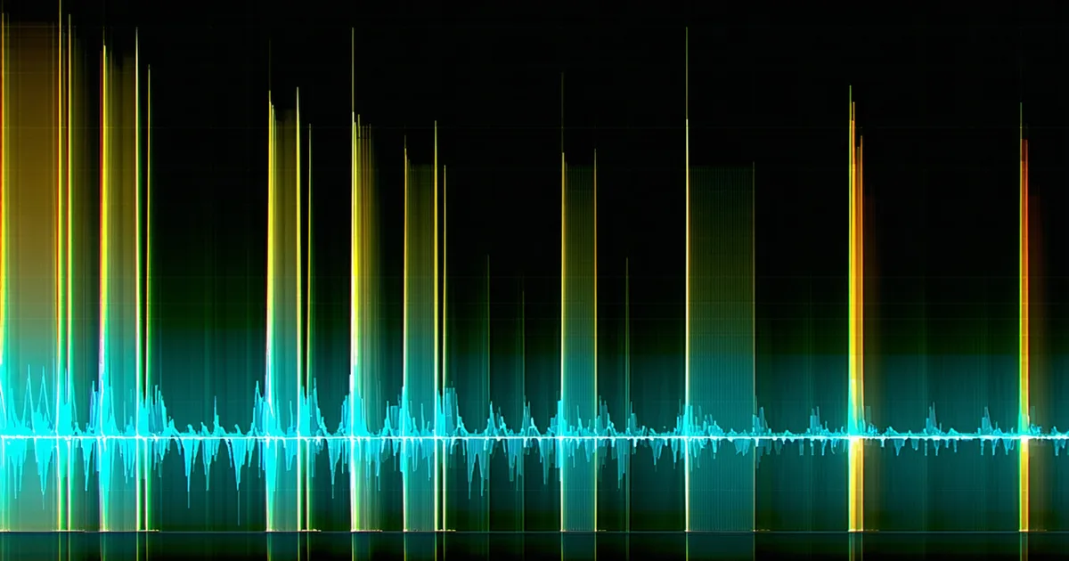
The standard preprocessing prescription for motor-imagery EEG is a bandpass filter in the 8–30 Hz range, targeting the mu (8–13 Hz) and beta (13–30 Hz) bands where event-related desynchronization (ERD) and post-movement beta rebound (ERS) are expressed. Bandpass filtering eliminates most power line interference and removes slow drift below 1 Hz. What it does not remove are artifacts that occupy the same frequency range as the signal of interest.
EMG contamination from neck and facial muscles — which is essentially unavoidable when a participant is in an exoskeleton harness applying effort — contains broadband power that bleeds into the beta band. Saccade-related EOG artifacts produce frontal contamination that projects posteriorly via volume conduction. Even swallowing generates a brief high-amplitude event that a bandpass filter passes through nearly intact.
Independent component analysis (ICA) can remove many of these sources, but ICA requires stationarity assumptions that the signal violates on the timescale of a clinical session. Online artifact rejection via ICA requires continuous model updating — which means you are no longer applying a fixed preprocessing transform to your data, you are applying an adaptive one, and that has implications for how you calibrate and validate the downstream classifier.
Laplacian referencing and common average referencing (CAR) both attenuate volume-conducted artifacts by emphasizing local sources. Laplacian referencing in particular improves spatial specificity around motor areas and reduces the influence of far-field noise. But referencing strategies alone cannot address non-stationary artifact sources, because the projection from artifact source to electrode changes as the artifact source (e.g., muscle group) changes its activity.
Session-to-Session Non-Stationarity in Feature Space
Even if we hold the electrode-scalp interface perfectly stable and eliminate all artifacts — which we cannot, but hypothetically — we would still face a fundamental problem: the neural signal itself changes between sessions.
Motor imagery ERD is not a fixed spectral signature. The frequency, spatial extent, and amplitude of mu-band desynchronization during imagined hand movement varies with fatigue, arousal, medication state, and the cumulative effect of neuroplastic changes from prior rehabilitation sessions. A participant early in rehabilitation therapy has a different motor-imagery EEG profile than the same participant eight weeks later — ideally because the therapy is working and cortical representation is reorganizing.
This means that a classifier trained at session one is approximating a decision boundary in feature space that will have shifted by session two. In Euclidean feature space (e.g., log-variance of CSP-filtered channels), this manifests as a translation and scaling of the class-conditional distributions. In covariance matrix space — where Riemannian geometry classifiers operate — it manifests as a change in the covariance structure of the data, detectable as a shift in the geometric mean of the SPD (symmetric positive definite) matrix manifold.
The magnitude of this shift is not trivial. Published benchmarks on openly available BCI datasets consistently show inter-session accuracy drops of 10–20 percentage points for static classifiers on 2-class motor imagery tasks. That is the difference between a clinically useful decoder and a coin flip at the lower end of the distribution.
Why Research Results Don't Transfer to Clinical Workflows
Academic BCI literature overwhelmingly reports results under controlled conditions: single-session or within-session cross-validation, fixed electrode placement by trained researchers, short recording durations (typically 30–90 minutes), and participants selected for clean, high-amplitude motor-imagery signals. These conditions are appropriate for demonstrating the theoretical capability of a signal processing method. They are not representative of a clinical deployment scenario.
In a neurorehabilitation clinic, EEG setup is performed by rehabilitation staff — not signal processing PhDs. Sessions may run two hours or longer. Participants have varying levels of neurological impairment that affect signal amplitude and spatial consistency. The same participant is seen across weeks and months of therapy. And critically, the clinical staff do not have the time or expertise to re-run calibration protocols and re-tune classifiers between every session.
Consider what happens in a realistic rehabilitation exoskeleton deployment: a participant arrives for their Monday session. The EEG cap is fitted by a rehabilitation technician following a standard protocol, but electrode placement has natural variability of ±5–10mm from the prior session. Impedances are checked and gelled. A two-minute calibration recording is taken. The decoder must now generalize from this calibration data — which reflects today's electrode configuration, today's participant state, and today's artifact environment — to the next 90 minutes of therapy.
Raw EEG decoded with a static pre-trained classifier will fail this scenario reliably. The question is not whether non-stationarity is a problem — it is. The question is which combination of alignment, re-referencing, adaptive covariance updating, and domain adaptation strategies keeps classification accuracy within clinically acceptable bounds across that 90-minute session and across the subsequent sessions.
What the Signal Pipeline Must Actually Handle
Designing a clinical BCI signal pipeline requires explicitly addressing each of the failure modes above, not assuming they will average out:
- Electrode drift compensation: Re-referencing strategies (CAR, Laplacian) reduce the effect of slow impedance drift. Online covariance updating — adapting the spatial filter or classifier covariance estimate as new data arrives — handles larger electrode displacements. Euclidean alignment between sessions re-centers the covariance matrix distribution before classification.
- Artifact handling: Band-specific artifact rejection (muscle contamination detection in the 20–40 Hz range) and online ICA or artifact subspace projection provide continuous cleaning without requiring a fixed preprocessing model.
- Session-to-session alignment: Some form of domain adaptation is required at session boundaries. This can be as lightweight as Euclidean alignment (re-centering each session's covariance matrices relative to the identity before Riemannian classification) or as complex as a full Riemannian transfer learning procedure. The choice depends on available calibration data, computational budget, and regulatory documentation requirements.
- Real-time latency constraints: All of this processing must complete within the latency budget. For rehabilitation feedback, the practical ceiling is approximately 100ms from motor intent to actuator command. Adaptive algorithms that require offline batch processing cannot be used for the online decode path.
The clinical deployment problem is fundamentally a systems engineering problem, not purely an algorithm design problem. Getting ERD classification to work in a controlled experiment requires a good algorithm. Getting it to work reliably in a clinic on a Thursday afternoon after two hours of therapy, with an electrode cap that has shifted since the morning calibration, requires an end-to-end pipeline designed around the failure modes that raw EEG encounters by default.
That pipeline design — rather than any individual classification innovation — is what separates research-grade BCI from clinical-grade BCI. And it is what the Synaptiq SDK was built to provide to device manufacturers who do not want to rebuild it themselves.