Any serious work on motor-imagery BCI decoding starts with a precise understanding of what you are actually trying to detect in the EEG signal. The terms "mu rhythm," "beta rebound," ERD, and ERS get used frequently, sometimes interchangeably and sometimes imprecisely. For clinical rehabilitation applications, these distinctions matter — both for designing the signal processing pipeline and for understanding what a classifier's output is actually measuring about the patient's neural state.
This is not a comprehensive review of the neurophysiology literature. It is a focused account of the specific oscillatory phenomena that the Synaptiq decoder targets, explained in enough depth that a device engineer integrating our SDK understands what the classifier is sensitive to, what conditions degrade it, and why the temporal and spectral structure of the features we extract is designed the way it is.
Event-Related Desynchronization: The Motor Imagery Signal
During voluntary movement or motor imagery, cortical oscillations in the mu band (approximately 8–13 Hz) and beta band (approximately 13–30 Hz) show a characteristic decrease in power over the sensorimotor cortex contralateral to the moved or imagined limb. This power decrease is event-related desynchronization (ERD).
The term "desynchronization" refers to the working hypothesis about the underlying neural mechanism: synchronized oscillatory activity in a cortical population produces a large-amplitude, rhythmic EEG signal; when the population is activated (during motor planning and execution), individual neurons fire in less synchronous patterns, reducing the collective oscillatory amplitude detectable at the scalp. This is sometimes described as an "event-related decrease in idle rhythms" — the motor cortex, when engaged, stops its idle oscillatory activity.
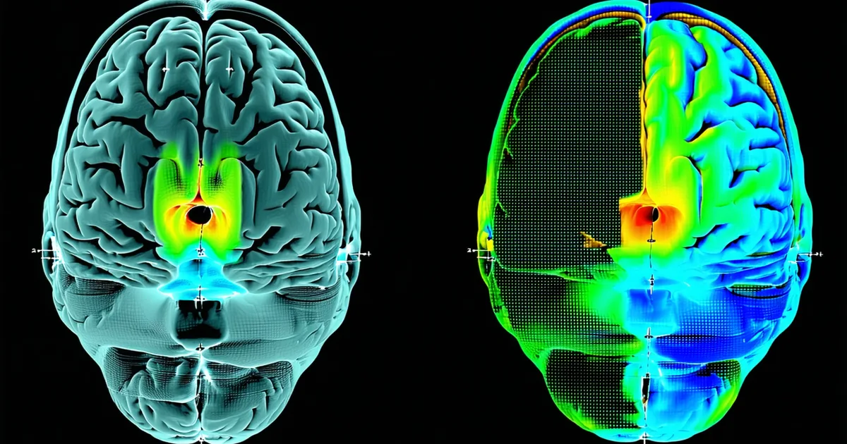
ERD onset typically precedes movement or motor imagery onset by 0.5–1.5 seconds, peaks around movement onset, and is expressed as a lateralized pattern: left-hand imagery produces right-hemisphere ERD (over electrode positions C4, CP4 in the 10-20 system) while right-hand imagery produces left-hemisphere ERD (over C3, CP3). This lateralization is the primary spatial discriminative feature that CSP spatial filters exploit.
The magnitude of ERD detectable at the scalp is highly variable. Well-practiced motor imagers show strong, focal mu-band ERD with clear lateralization. Participants early in motor imagery training — a realistic clinical scenario for stroke rehabilitation patients who may never have used BCI before — show weaker, more diffuse signals that are harder to classify. This is one reason why calibration session length and trial count matter so much in clinical BCI: you need enough labeled data to estimate the participant's specific ERD pattern accurately, and that estimate is more noisy for participants with weaker signals.
Beta Rebound: The Post-Movement Signature
After the conclusion of movement or motor imagery, there is a characteristic increase in beta power that overshoots the pre-movement baseline. This is event-related synchronization (ERS), specifically the beta rebound or post-movement beta synchronization (PMBS). It typically begins 300–700 ms after movement end, peaks around 1–2 seconds post-movement, and can persist for 2–3 seconds.
The beta rebound is thought to reflect an active motor inhibitory process — the reinstatement of the cortical idle state after motor engagement. Its amplitude and timing correlate with successful motor execution and may reflect aspects of sensorimotor integration distinct from the planning-related ERD. For rehabilitation applications, the beta rebound is relevant because it provides a temporal boundary marker: strong beta rebound after an imagined movement indicates that the cortical motor cycle has completed.
Clinically, several studies have suggested that the amplitude and timing of beta rebound may correlate with motor recovery outcomes — participants who show more pronounced beta dynamics during BCI-assisted rehabilitation tend to show better clinical improvement. We are careful not to make clinical outcome claims, but the physiological plausibility is well-established in the neuroscience literature, and it motivates our design choice to track both ERD amplitude and beta rebound timing in the Synaptiq feature set, rather than treating the post-movement window as uninformative.
Individual Frequency Variability and the Clinical Challenge
The frequency ranges given above — mu at 8–13 Hz, beta at 13–30 Hz — are population averages. Individual peak frequencies vary substantially. A participant's mu peak may be at 9.5 Hz, 10.5 Hz, or 12 Hz. Their beta ERD may be concentrated in the 14–20 Hz range or extend into the 24–28 Hz range. This individual variability is why a fixed 8–30 Hz bandpass filter is a reasonable starting point but not optimal for any given individual.
In rehabilitation populations, there is additional variability introduced by pathology. Following stroke, the affected hemisphere often shows altered oscillatory dynamics: reduced mu amplitude, shifted peak frequencies, and less distinct lateralization. This is partly why stroke rehabilitation is one of the motivating clinical applications for BCI — the damaged hemisphere's motor representations are being reinforced through neurofeedback — but it also means the signal is weaker and less discriminative, requiring more careful preprocessing and classifier adaptation.
From a signal processing standpoint, individual frequency variability motivates the filter bank approach (FBCSP or a Riemannian equivalent): by decomposing the signal into multiple sub-bands and selecting or weighting features from each, the decoder can adapt to individual frequency profiles without explicit individual calibration of band limits. This is one reason we use a multi-band approach in the Synaptiq pipeline rather than relying on a single fixed bandpass — the right frequency range for a 45-year-old stroke patient with right hemisphere damage is different from the right range for a 30-year-old with an acute traumatic brain injury, and we do not want the engineer integrating the SDK to need to tune this per patient.
Spectral Features vs. Amplitude-Only Approaches
A common early approach to motor imagery classification uses bandpass-filtered amplitude or log-power in a fixed frequency band as the primary feature. This captures the dominant ERD effect but misses important information about the spectral structure of the signal.
Covariance matrix-based features — which encode the inter-channel and intra-channel spectral relationships simultaneously — capture more of the available discriminative structure. A covariance matrix computed from a bandpass-filtered multichannel EEG segment encodes both the power in each channel (diagonal elements) and the phase relationships between channels (off-diagonal elements). The phase relationships carry information about the spatial coherence of motor-imagery oscillations that amplitude-only features discard.
This is one of the reasons why Riemannian geometry classifiers operating on covariance matrices consistently outperform classifiers operating on scalar power features, particularly in cross-session evaluation: the covariance structure encodes richer information about the neural state, and the Riemannian metric provides a distance measure that is sensitive to this structure in a geometrically appropriate way.
Implications for Decoder Design in Rehabilitation Applications
Understanding the ERD/ERS physiology clarifies several design choices in clinical BCI:
Trial window selection: The ERD is most pronounced 0.5–2 seconds after motor imagery onset. A feature extraction window of 0.5–2.5 seconds (with onset offset by 0.5–1 seconds from the cue) captures the peak discriminative signal for most participants. Shorter windows (250ms) sacrifice accuracy for latency; longer windows (3+ seconds) add latency without proportional accuracy gain for most participants. The right choice depends on the latency budget of the specific device and therapy protocol.
Rest vs. imagery classification: Many clinical protocols require detecting when a participant is attempting motor imagery versus resting. ERD provides a reliable signal for this, but the rest-state mu rhythm amplitude is itself variable — it is the "idle rhythm" and fluctuates with arousal and attention. Thresholding on absolute ERD magnitude is unreliable; relative change from a participant-specific rest baseline is more consistent.
Session length effects: ERD amplitude decreases with fatigue over a long session. The beta rebound amplitude may also change. These changes in signal magnitude are partly what drives within-session classification accuracy degradation in naive static decoders — the signal the classifier was trained to recognize becomes weaker and noisier as the session progresses. Online covariance updating that adapts to these amplitude changes is therefore not just handling electrode drift; it is tracking the changing physiological state of the participant's motor cortex.
The rehabilitation-specific conclusion is this: a reliable motor-imagery decoder for neurorehabilitation must track spectral dynamics rather than just spatial amplitude. The mu and beta bands are the right frequency targets, but their individual expression varies enough across participants, sessions, and fatigue levels that a static model designed around population averages will fail a substantial fraction of participants in the clinic. Adaptive, individual-specific decoding is not a research luxury — for rehabilitation BCI, it is the minimum requirement for consistent clinical utility.